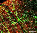
File:PLoSBiol4.e126.Fig6fNeuron.jpg
外觀

預覽大小:687 × 600 像素。 其他解析度:275 × 240 像素 | 550 × 480 像素 | 915 × 799 像素。
原始檔案 (915 × 799 像素,檔案大小:787 KB,MIME 類型:image/jpeg)
檔案歷史
點選日期/時間以檢視該時間的檔案版本。
| 日期/時間 | 縮圖 | 尺寸 | 使用者 | 備註 | |
|---|---|---|---|---|---|
| 目前 | 2013年2月13日 (三) 10:34 |  | 915 × 799(787 KB) | Hic et nunc | Maßstab wieder rein |
| 2013年2月13日 (三) 07:17 |  | 921 × 805(836 KB) | Hic et nunc | watermark removed | |
| 2008年1月31日 (四) 21:30 |  | 922 × 806(804 KB) | Dietzel65 | {{Information |Description={en|After the original figure legend: Coronal section containing the chronically imaged pyramidal neuron “dow” (visualized by green GFP) does not stain for GABA (visualized by antibody staining in red). Confocal image stack, |
檔案用途
下列頁面有用到此檔案:
全域檔案使用狀況
以下其他 wiki 使用了這個檔案:
- als.wikipedia.org 的使用狀況
- ar.wikipedia.org 的使用狀況
- arz.wikipedia.org 的使用狀況
- as.wikipedia.org 的使用狀況
- azb.wikipedia.org 的使用狀況
- ca.wikipedia.org 的使用狀況
- cs.wikipedia.org 的使用狀況
- cy.wikipedia.org 的使用狀況
- de.wikipedia.org 的使用狀況
- de.wikibooks.org 的使用狀況
- Natur und Technik für den Pflichtschulabschluss: Das Leben
- Natur und Technik für den Pflichtschulabschluss: Die Evolution der Zelle
- Natur und Technik für den Pflichtschulabschluss: Neuron
- Natur und Technik für den Pflichtschulabschluss: Menschliche Gewebe
- Benutzer:Yomomo/ NuT
- Natur und Technik für den Pflichtschulabschluss/ Buch
- de.wikiversity.org 的使用狀況
- de.wiktionary.org 的使用狀況
- en.wikipedia.org 的使用狀況
- en.wikiquote.org 的使用狀況
- es.wikipedia.org 的使用狀況
- es.wikibooks.org 的使用狀況
- eu.wikipedia.org 的使用狀況
- fa.wikipedia.org 的使用狀況
- fr.wikipedia.org 的使用狀況
- fr.wikiversity.org 的使用狀況
- ga.wikipedia.org 的使用狀況
- gd.wikipedia.org 的使用狀況
- gl.wikipedia.org 的使用狀況
- he.wikipedia.org 的使用狀況
- hi.wikipedia.org 的使用狀況
- hy.wikipedia.org 的使用狀況
- ka.wikipedia.org 的使用狀況
- kn.wikipedia.org 的使用狀況
- ko.wikipedia.org 的使用狀況
檢視此檔案的更多全域使用狀況。


